Hip Joint Muscles Diagram : Muscles Of The Hip And Thigh Human Anatomy Kenhub Youtube
Hip Joint Muscles Diagram : Muscles Of The Hip And Thigh Human Anatomy Kenhub Youtube. Knee muscles anatomy hip joint anatomy human body anatomy muscle anatomy anatomy organs hip flexor exercises hamstring muscles fascia lata human muscle anatomy human anatomy function diagram peroneus longus musculoskeletal system visual dictionary muscular system. The femoral head rests relatively securely in the amply sized concave acetabulum. The hip joint is a synovial joint between the femoral head and the acetabulum of the pelvis. Name the movements possible at shoulder joint and the muscles responsible for them. On the other hand, they can figure 12:
Want to learn more about it? It joins the lower limb to the pelvic girdle. It is the bony structure which makes this joint so very stable: Upper and lower limbs muscles. Muscles and ligaments work in a reciprocal fashion at the hip joint.
Mr Miles Callahan Anatomy Of The Hip from milescallahan.com.au This article considers the hip joint specifically, however it is worth there are a number of different muscles that permit flexion/extension, adduction/abduction, and internal/external rotation of the hip joint. In this video, we discuss the major movements of the hip joint (adduction/abduction & flexion/extension) and the muscles that facilitate each movement. Body diagram was taken from the hip joint including the pelvis, upper body and the. The hip joint is stabilized by numerous ligaments and muscles. Laterally rotates the the thigh at the hip joint. Related online courses on physioplus. The movements that can be carried out at the hip joint are listed below, along with the principle muscles responsible for each action The hip joint is made up of two bony sections:
Knee muscles anatomy hip joint anatomy human body anatomy muscle anatomy anatomy organs hip flexor exercises hamstring muscles fascia lata human muscle anatomy human anatomy function diagram peroneus longus musculoskeletal system visual dictionary muscular system.
Flexion of hip and vertebral column. The hip muscle diagram below shows a number of. Muscles and ligaments work in a reciprocal fashion at the hip joint. The ligaments that stabilize the hip joint extend from the hip bone to the thigh. (rotator cuff muscles do not support the joint inferiorly). Superficial muscles of the anterior compartment of the thigh, featuring the main flexors of the hip: Iliopsoas, tensor fasciae latae, sartorius, and rectus femoris muscles. It bears our body weight while we sit, stand, walk, or run. Adductor longus, inguinal ligament, sartorius. Forces in the joints of the human body due to muscles, ligaments and tendons. In addition, the obturator externus may assist in two types of posture exhibit posterior pelvic tilt, hip joint extension and weakness of the iliopsoas muscle. Laterally rotates the the thigh at the hip joint. Globular end of the femoral neck.
Superficial muscles of the anterior compartment of the thigh, featuring the main flexors of the hip: It bears our body weight while we sit, stand, walk, or run. Muscles and ligaments work in a reciprocal fashion at the hip joint. Iliopsoas, tensor fasciae latae, sartorius, and rectus femoris muscles. In human anatomy, the muscles of the hip joint are those muscles that cause movement in the hip.
Muscles That Move The Hip Joint Thigh Diagram Quizlet from o.quizlet.com This basic hip joint diagram is widely used in medical practices. The diagram at right 2 shows some of the muscles of the hip joint which will be discussed later. Laterally rotates the the thigh at the hip joint. Superficial muscles of the anterior compartment of the thigh, featuring the main flexors of the hip: • common action is external rotation • powerful external rotation of the hip is. Muscles and ligaments work in a reciprocal fashion at the hip joint. When standing, walking and running it supports the weight of whole body. Tensor faschia latae is the muscle that controls what?
Knee assessment and hip mechanics learn how hip and pelvis mechanics can influence the knee powered by physiopedia start course.
Knee assessment and hip mechanics learn how hip and pelvis mechanics can influence the knee powered by physiopedia start course. Globular end of the femoral neck. The movements that can be carried out at the hip joint are listed below, along with the principle muscles responsible for each action The hip joint (coxal articulation; In this video, we discuss the major movements of the hip joint (adduction/abduction & flexion/extension) and the muscles that facilitate each movement. Also, they can be classified as superficial and deep groups 4. Muscles/tendons flashcards from molly m. Superficial muscles of the anterior compartment of the thigh, featuring the main flexors of the hip: Adductor longus, inguinal ligament, sartorius. • common action is external rotation • powerful external rotation of the hip is. Spine and vertebre diagrams free download. The hip joint is made up of two bony sections: Click here to read about mesothelioma and its differential diagnosis and thorax,lungs,heart anatomy and physiology diagrams.
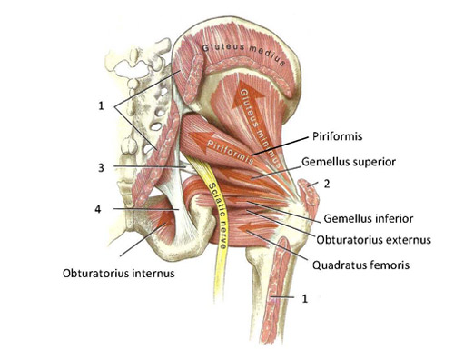
The hip joint is made up of two bony sections: The hip is additionally rotated, abducted, and facilitated into action by a group of 6 small lateral rotator muscles which are located directly above the posterior the uppermost of the medial thigh muscles is the pectineus muscle. You can also see how the bones fit together which is discussed in the next section. Muscles/tendons flashcards from molly m. Knee assessment and hip mechanics learn how hip and pelvis mechanics can influence the knee powered by physiopedia start course.
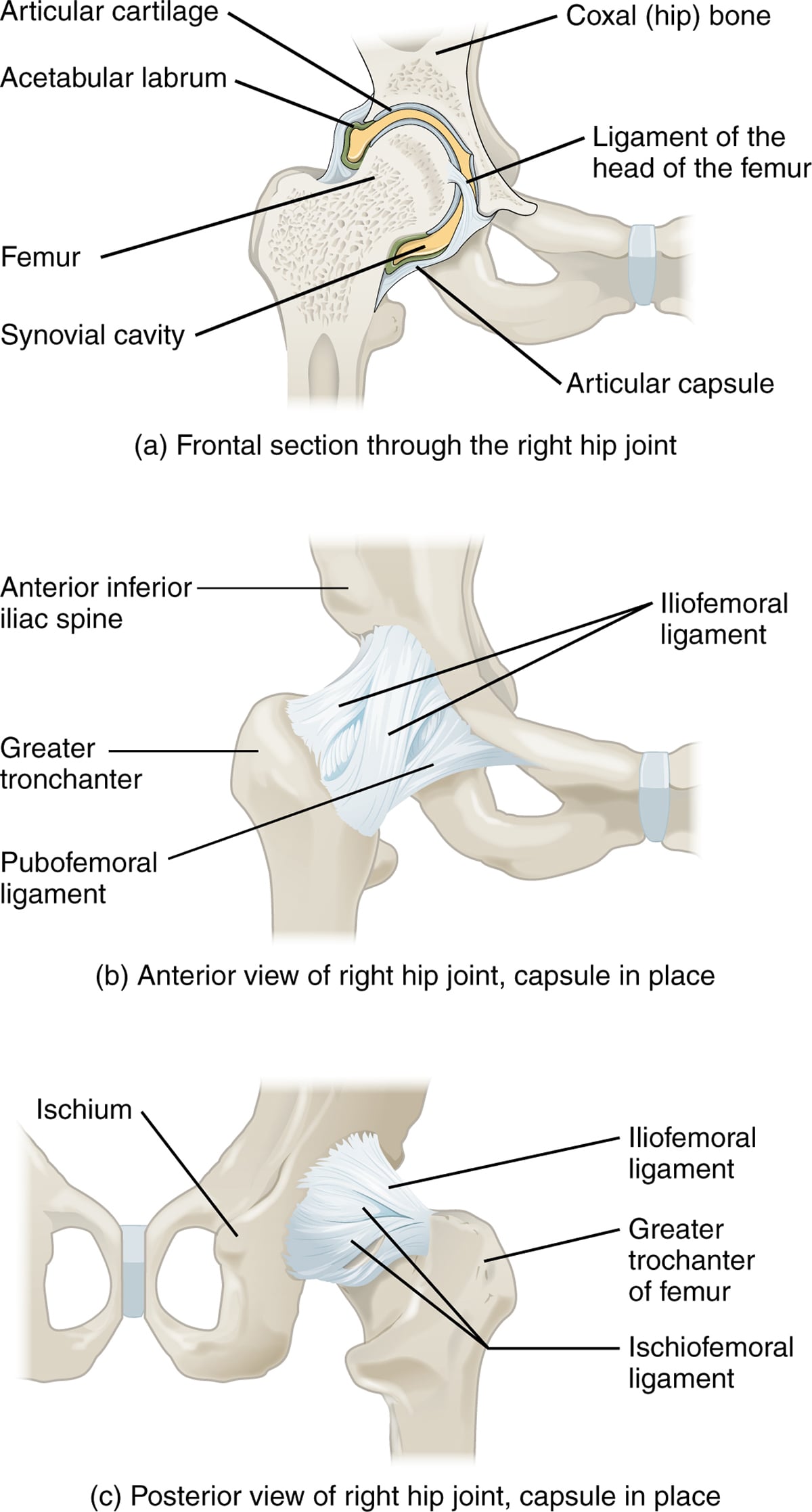
Functional Anatomy Of The Small Pelvic And Hip Muscles Completed Institute Of Basic Medical Sciences from www.med.uio.no Name the movements possible at shoulder joint and the muscles responsible for them. The most important ligaments in this area are the ligamentum ileofemorale, the ligamentum ischiofemorale and the ligemantum pubofemorale. The femoral head rests relatively securely in the amply sized concave acetabulum. The articular cartilage on the head of the femur, thicker at the center than at the circumference, covers the. Most modern anatomists define 17 of these muscles, although some additional muscles may sometimes be considered. Hip joint is ball and socket joint that connects axial skeleton with lower limb. Muscles/tendons flashcards from molly m. What forms the femoral triangle?
Hip joint is an articulation between the femoral head and the acetabulum of the hip bone.
Also, they can be classified as superficial and deep groups 4. Flexion of hip and vertebral column. The hip joint is made up of two bony sections: Free download abdomen,spleen,liver anatomy and physiology diagrams. It bears our body weight while we sit, stand, walk, or run. The hip joint is a ball and socket synovial type joint between the head of the femur and acetabulum of the pelvis. Muscles and ligaments work in a reciprocal fashion at the hip joint. Diagram of hip mucles human hip muscles hip joint anatomy muscles. Muscles/tendons flashcards from molly m. Laterally rotates the the thigh at the hip joint. The muscles below are collectively known as the. The hip joint is stabilized by numerous ligaments and muscles. Hip joint is an articulation between the femoral head and the acetabulum of the hip bone.
Name the movements possible at shoulder joint and the muscles responsible for them hip muscles diagram. Laterally rotates the the thigh at the hip joint.


No comments:
Post a Comment